
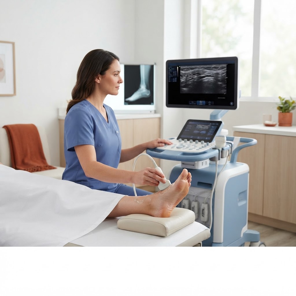
The Role of Imaging in Evaluating Chronic Plantar Fasciitis: Diagnostic Paths
Persistent heel pain is a common frustration that can significantly impact your daily mobility. While many cases of plantar fasciitis respond well to rest, stretching, and orthotic inserts, some individuals experience symptoms that linger despite consistent home care. When pain persists for several months, it may be time to seek a formal diagnostic evaluation to rule out other pathology or confirm the severity of the fascial inflammation.
When to Seek Professional Imaging
Not every heel ache requires medical imaging. However, if you have faithfully followed a conservative treatment plan for six to eight weeks without improvement, or if you notice significant swelling, inability to bear weight, or numbness in the foot, you should consider a clinical review. Imaging provides clarity that physical exams alone cannot, allowing you to visualize the exact structures of the foot.
X-Rays vs. Ultrasound: Choosing the Right Modality
When you approach a provider to discuss imaging, it is important to understand the difference between the two most common tools for foot pain.
Weight-Bearing X-Rays
X-rays are excellent for evaluating bone structure. If your provider suspects a stress fracture, bone cyst, or wants to assess the presence of a large calcaneal spur, a weight-bearing X-ray is the gold standard. It captures the foot while it is supporting your body weight, which can reveal alignment issues or bony protrusions that contribute to discomfort. However, X-rays are limited because they do not capture soft tissues like the plantar fascia.
Musculoskeletal (MSK) Ultrasound
When the concern is specifically soft-tissue health, ultrasound is often the superior choice. An MSK ultrasound can clearly measure the thickness of the plantar fascia. Typically, a fascia measuring over 4 millimeters in thickness is a clinical indicator of fasciitis. Ultrasound is dynamic, meaning a technician can apply pressure to the area to see how the tissue reacts in real time, often providing a more definitive look at inflammation or micro-tears than a static X-ray.
How to Prepare for Your Appointment
If your provider determines that an imaging study is necessary, preparation is minimal but important. For an MSK ultrasound, wear loose-fitting pants that can be rolled up well above the ankle. You do not need to fast or follow any dietary restrictions. Be prepared to discuss your pain history, including when it feels worst—usually, this is the first few steps in the morning—and any specific activities that trigger a flare-up.
Streamlining Your Care with Diagnostic Orders Direct
Obtaining a provider order for these studies can sometimes be the biggest hurdle. Whether you are uninsured, dealing with insurance coverage delays, or simply lack a primary care physician to facilitate these requests, Diagnostic Orders Direct (https://diagnosticordersdirect.com) provides a streamlined solution. We offer virtual provider reviews for diagnostic imaging requests, including ultrasounds and X-rays, for just $40. If medically appropriate, our licensed providers can issue the necessary orders, allowing you to schedule your imaging at a facility of your choice without unnecessary delays.
Taking the Next Step
Persistent heel pain does not have to be your permanent reality. By utilizing appropriate imaging to understand what is happening beneath the skin, you and your healthcare team can develop a targeted recovery plan. When you are ready to initiate the process, our team is here to help you get the documentation you need.
Important
Diagnostic Orders Direct is not an imaging center, laboratory, radiology service, or emergency service. If you are experiencing a medical emergency, please call 911 or visit the nearest emergency room immediately. Orders are not guaranteed and are issued only when determined to be medically appropriate by a licensed provider. Our services are not a replacement for comprehensive primary care.